Descripción microscópica:
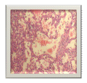
Imagen 2. Espacios vasculares recubiertos por células endoteliales atípicas.

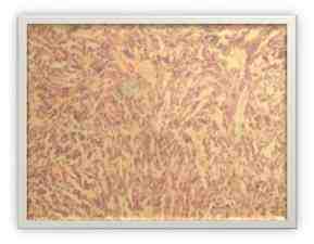
Imagen 3. Aspecto fusocelular, formación de estructuras vasculares.

Referencias bibliográficas.
- Marshall A. Lichtman, Thomas J. Kipps, Uri Seligsohn, Kenneth Kaushansky, Josef T. Prchal. Williams Hematology, 8th Ed. Beiing: McGraw-Hill; 2010
- González González Vicente Raúl, Céspedes González Reina, Capote Pereira Lázaro, Pérez Bomboust Isela, Arpa Gámez Ángel. Angiomatosis difusa del bazo como causa de esplenomegalia. Rev Cub Med Mil [Internet]. 2008 Jun [citado 2013 Mar 29]; 37(2): Disponible en: http://scielo.sld.cu/scielo.php?script=sci_arttext&pid=S0138-65572008000200008&lng=es.
- Capote Pereira Lázaro L., Carral Novo Juan M., Pérez Bomboust Isela, Capote Leiva Eliseo, Rebollar Martínez Alberto, Rodríguez Apolinario Norlan et al. Ruptura esplénica espontánea postrasplante y angiomatosis difusa del bazo. Rev Cub Med Mil [Internet]. 2006 Sep [citado 2013 Mar 29]; 35(3): Disponible en: http://scielo.sld.cu/scielo.php?script=sci_arttext&pid=S0138-65572006000300011&lng=es.
- Wang HL. Li KW. Wang J. Sclerosing angiomatoid nodular transformation of the spleen: report of five cases and review of literature. Chin Med J (Engl). 2012; 125(13):2386-9
- WANG Z. Yu Z. Su Y. Yang H. Cao L. Zhao X. et al. Kasabach-Merritt syndrome caused by giant hemangiomas of the spleen in patients with Proteus syndrome. Blood Coagul Fibrinolysis. 2007; 18(5):505-8
- Min KW, Reed JA, Welch DF et al. Morphologically variable bacilli of catscratch disease identified by immunocytochemical labeling with antibodies to Rochalimaea Henselae. Am. J. Clin. Pathol. 1994;101:607-610.
- Stevens DL, Bisno AL, Chambers HF, Everett ED, Dellinger P, Goldstein EJ, et al. Practice guidelines for the diagnosis and management of skin and soft-tissue infections. Clin Infect Dis. Nov 15 2005;41(10):1373-406..
- Kim HJ. Kim KW. Yu ES. Byun JH. Lee SS. Kim JH. ET AL. Sclerosing angiomatoid nodular transformation of the spleen: clinical and radiologic characteristics. Acta Radiol. 2012; 53(7):701-6
- Thacker C. Korn R. Millstine J Harvin H. Van Lier Ribbink JA. Gotway MB. Sclerosing angiomatoid nodular transformation of the spleen: CT, MR, PET, and ⁹⁹(m)Tc-sulfur colloid SPECT CT findings with gross and histopathological correlation. Abdom Imaging. 2010; 35(6):683-9
- Cao JY. Zhang H. Wang WP. Ultrasonography of sclerosing angiomatoid nodular transformation in the spleen. World J Gastroenterol. 2010; 16(29):3727-30
- Kornprat P. Beham-Schmid C. Parvizi M. Portugaller H. Bernhardt G, Mischinger HJ. Incidental finding of sclerosing angiomatoid nodular transformation of the spleen. Wien Klin Wochenschr. 2012; 124(3-4):100-3